
Technology and safety
The equipment and safety steps used on every patient, and what each one means for you.
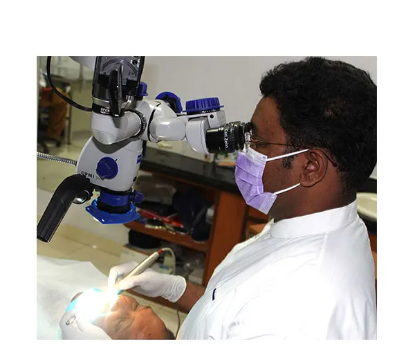
Surgical operating microscope
Dr. Vinod was an early adopter of the surgical operating microscope from Carl Zeiss, Germany, for root canal treatment in India. It lights and magnifies the tooth so he can diagnose and treat what the naked eye misses, including hidden canals, cracks and broken instruments.
That makes it possible to treat difficult cases that would otherwise end in extraction. The microscope also records images, so you can see what was found.
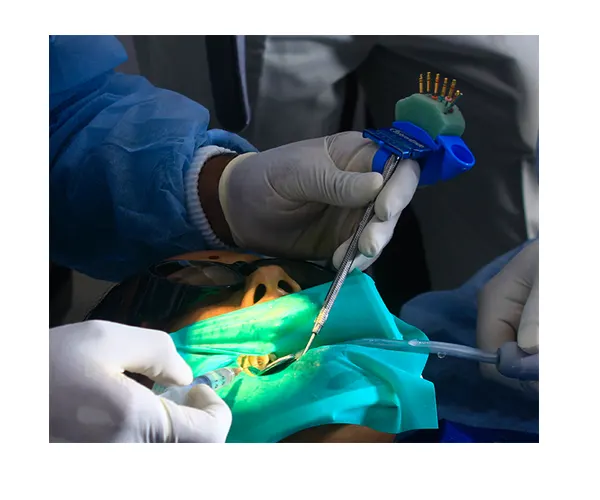
A rubber dam on every case
A thin sheet that isolates the tooth being treated.
- Keeps saliva out. Saliva carries germs into the root canal, which can slow healing and lead to failure.
- Allows strong disinfecting solutions to be used safely inside the canal.
- Stops you from swallowing or breathing in small instruments.
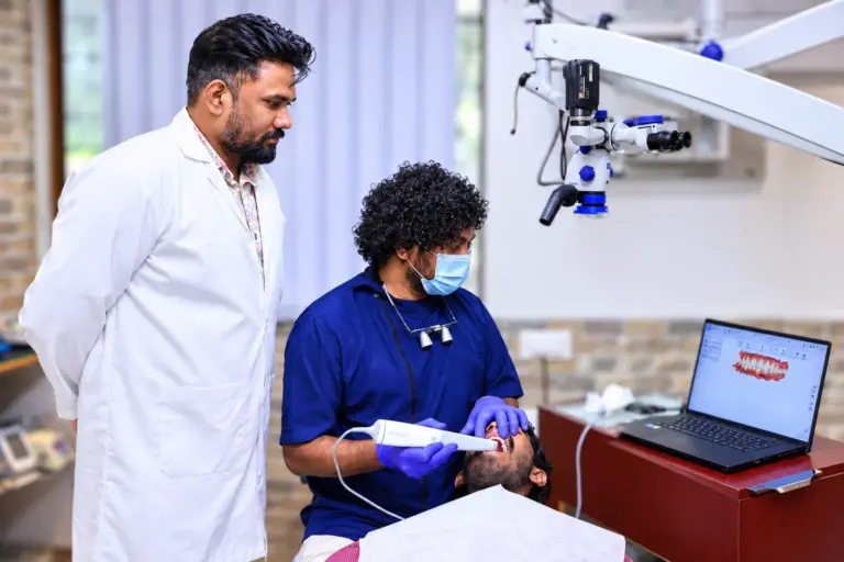
Digital scanning
A hand-held intraoral scanner records the shape of your tooth in a few minutes, without the bulky impression trays and messy impression material. The digital scan is used for crowns, inlays and onlays, and it is saved with your records so it can be compared at your yearly check.
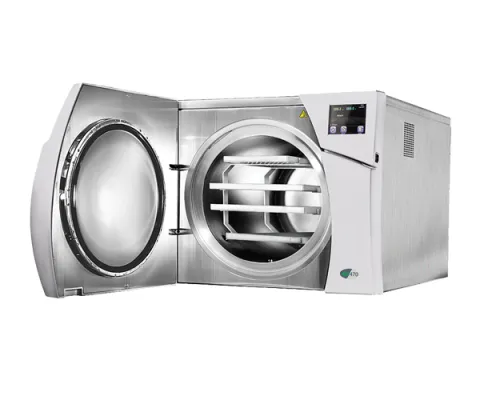
Sterilisation
Every instrument goes through three stages: disinfection, ultrasonic cleaning and autoclaving. Files, burs and other consumables are single-use.
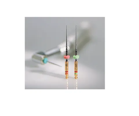
Rotary nickel titanium files
Flexible instruments that resist breaking and clean and shape curved canals well. They shorten treatment time and help in complicated cases.
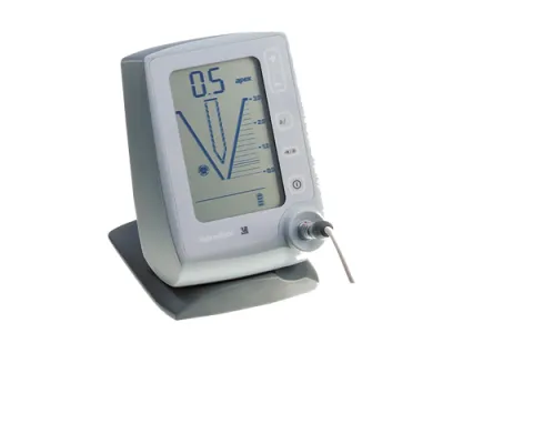
Apex locator
An electronic device that measures the length of each root canal, so it is cleaned and sealed right to its end.
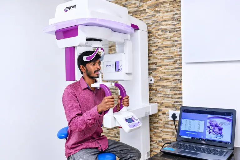
Digital X-rays
Digital sensors from Schick replace photographic film, and a digital full-mouth X-ray (OPG) captures every tooth and both jaws in a single image. The clinic’s figure for the reduction in radiation exposure is up to 90 percent, and images can be stored and shared with you instantly.